Decoding Twin Ultrasound Images: A Comprehensive Guide
The joy of expecting a child is often amplified when parents learn they are expecting twins. The journey begins with the confirmation, frequently revealed through twin ultrasound images. Understanding these images can be both exciting and a little daunting. This comprehensive guide aims to demystify twin ultrasound images, providing expecting parents with the knowledge to interpret what they see and ask informed questions during their prenatal appointments.
Understanding the Basics of Ultrasound Technology
Before diving into the specifics of twin ultrasound images, it’s crucial to understand the underlying technology. Ultrasound, also known as sonography, uses high-frequency sound waves to create real-time images of internal body structures. These sound waves are emitted by a transducer, which is moved over the abdomen. The waves bounce back differently depending on the density of the tissue they encounter, and these echoes are then processed into an image.
In obstetrics, ultrasound is a cornerstone of prenatal care. It’s used to confirm pregnancy, estimate gestational age, assess fetal growth, and, of course, determine if there are multiple fetuses present. The clarity and detail of twin ultrasound images have improved dramatically over the years, allowing for earlier and more accurate diagnoses.
Identifying Twins on an Ultrasound
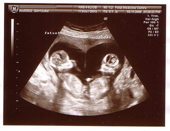
The moment of discovery – seeing two distinct gestational sacs or fetal poles on the screen – is often a memorable one. The earliest twin ultrasound images, typically taken around 6-8 weeks of gestation, may show two separate gestational sacs. As the pregnancy progresses, the images become clearer, revealing more details about each baby.
Key things to look for in twin ultrasound images include:
- Gestational Sacs: Two separate sacs indicate a dichorionic-diamniotic (Di-Di) twin pregnancy, meaning each twin has its own chorion (outer membrane) and amnion (inner membrane).
- Fetal Poles: These are the earliest visible forms of the developing embryos. Seeing two fetal poles confirms the presence of twins.
- Heartbeats: The presence of two distinct heartbeats is a definitive sign of a twin pregnancy.
- Amniotic Membranes: In monochorionic pregnancies (where twins share a chorion), the number of amniotic membranes (one or two) determines whether it’s a monochorionic-diamniotic (Mo-Di) or monochorionic-monoamniotic (Mo-Mo) pregnancy.
Types of Twin Pregnancies and Their Ultrasound Appearances
Twin pregnancies are classified based on chorionicity and amnionicity, which refer to the number of chorions and amnions each twin has. This classification is vital because it affects the risks and management of the pregnancy. Understanding these distinctions can help you better interpret your twin ultrasound images.
Dichorionic-Diamniotic (Di-Di) Twins
Di-Di twins each have their own chorion and amnion. This is the most common type of twin pregnancy and is always the case for dizygotic (fraternal) twins. In twin ultrasound images, Di-Di twins are characterized by:
- Two separate gestational sacs in early pregnancy.
- A thick dividing membrane between the twins, composed of two chorions and two amnions.
- The “lambda” or “twin peak” sign, a triangular projection of placental tissue extending between the layers of the dividing membrane. This sign is highly suggestive of a Di-Di pregnancy.
Monochorionic-Diamniotic (Mo-Di) Twins
Mo-Di twins share a chorion but have separate amniotic sacs. This type of twin pregnancy occurs in about 70% of monozygotic (identical) twin pregnancies. In twin ultrasound images, Mo-Di twins are identified by:
- A single chorionic sac surrounding both twins.
- A thin dividing membrane between the twins, composed of two amnions.
- The absence of the “lambda” or “twin peak” sign.
Monochorionic-Monoamniotic (Mo-Mo) Twins
Mo-Mo twins share both a chorion and an amnion. This is the rarest and highest-risk type of twin pregnancy, occurring in only about 1% of monozygotic twin pregnancies. In twin ultrasound images, Mo-Mo twins are characterized by:
- A single gestational sac with no dividing membrane.
- The presence of two fetuses within the same amniotic sac.
- The potential for cord entanglement, which can be visualized on ultrasound.
What to Expect During a Twin Ultrasound
A typical twin ultrasound examination is similar to a singleton pregnancy ultrasound, but it may take longer to ensure that both babies are thoroughly assessed. The sonographer will measure each baby’s crown-rump length (CRL) in the first trimester to estimate gestational age. In later trimesters, they will measure the head circumference, abdominal circumference, and femur length of each twin to assess growth.
During the ultrasound, the sonographer will also evaluate:
- Placental Location: The position and structure of the placenta(s) are assessed to identify potential complications like placenta previa.
- Amniotic Fluid Levels: The amount of amniotic fluid surrounding each twin is measured to detect oligohydramnios (low fluid) or polyhydramnios (excess fluid).
- Fetal Anatomy: A detailed anatomical survey is performed to screen for any structural abnormalities in each twin.
- Umbilical Cord Doppler Studies: Blood flow through the umbilical cords is assessed to ensure that each twin is receiving adequate nutrients and oxygen.
Common Concerns and Questions About Twin Ultrasounds
Many expecting parents have questions and concerns about twin ultrasounds. Here are some common ones:
- Accuracy of Gestational Age: Ultrasound is generally accurate in estimating gestational age, especially in the first trimester. However, there can be slight variations in measurements between the twins.
- Detecting Twin-to-Twin Transfusion Syndrome (TTTS): TTTS is a serious complication that can occur in Mo-Di twin pregnancies. It involves an unequal sharing of blood supply between the twins. Ultrasounds are used to monitor amniotic fluid levels and blood flow in each twin to detect TTTS early.
- Growth Discordance: This refers to a significant difference in the size of the twins. Ultrasounds are used to monitor fetal growth and identify growth discordance, which may indicate problems with placental function or fetal health.
- Vanishing Twin Syndrome: In some cases, one of the twins may stop developing and be reabsorbed by the mother’s body. This is known as vanishing twin syndrome and can sometimes be seen on early twin ultrasound images.
Advanced Ultrasound Techniques for Twin Pregnancies
In addition to standard 2D ultrasound, advanced techniques like 3D and 4D ultrasound can provide more detailed views of the twins. 3D ultrasound creates static three-dimensional images, while 4D ultrasound adds the element of time, allowing for real-time visualization of fetal movements. These techniques can be particularly helpful in assessing facial features and identifying certain structural abnormalities.
Fetal echocardiography, a specialized ultrasound that focuses on the fetal heart, may be recommended for twin pregnancies, especially if there is a family history of heart defects. This allows for early detection of any cardiac abnormalities in the twins.
Interpreting Your Twin Ultrasound Report
After your twin ultrasound, you will receive a report that summarizes the findings. This report will include measurements of each twin, assessments of placental location and amniotic fluid levels, and any other relevant observations. It’s important to discuss the report with your healthcare provider to fully understand the implications of the findings.
Key things to look for in the report include:
- Chorionicity and Amnionicity: This will determine the type of twin pregnancy you have (Di-Di, Mo-Di, or Mo-Mo).
- Gestational Age: The estimated gestational age based on ultrasound measurements.
- Fetal Weights: The estimated weight of each twin.
- Amniotic Fluid Index (AFI): A measure of the amount of amniotic fluid surrounding each twin.
- Any Abnormalities: Any structural abnormalities or other findings that require further evaluation.
The Emotional Aspect of Seeing Twin Ultrasound Images
Beyond the medical information, twin ultrasound images often evoke a strong emotional response. Seeing two babies on the screen can be both overwhelming and incredibly joyful. It’s a moment that many parents cherish and remember for a lifetime. Sharing these images with family and friends can be a special way to announce the exciting news.
However, it’s also important to acknowledge that a twin pregnancy can come with increased anxiety and concerns. It’s crucial to have open and honest communication with your healthcare provider and to seek support from other twin parents. [See also: Support Groups for Twin Parents] Understanding your twin ultrasound images is a step towards feeling more informed and empowered throughout your pregnancy journey.
Conclusion: Empowering Parents Through Knowledge of Twin Ultrasound Images
Twin ultrasound images are a powerful tool in prenatal care, providing valuable information about the health and development of your babies. By understanding the basics of ultrasound technology, the different types of twin pregnancies, and what to expect during an ultrasound examination, you can feel more confident and informed throughout your pregnancy. Remember to ask questions, seek support, and cherish the incredible journey of expecting twins. The advancements in ultrasound technology continue to improve the detection and management of twin pregnancies, ensuring the best possible outcomes for both mothers and babies. The ability to visualize and monitor the growth of twins through twin ultrasound images is a testament to the progress of modern medicine and a source of hope and excitement for expecting parents.

